
Mitochondrial Genetics: Heteroplasmy, Inheritance, and Disease
By Sequencing Team, The team of bioinformaticians, Genetic Health Coaches, and writers at Sequencing.
Oct 5, 2026
Heteroplasmy
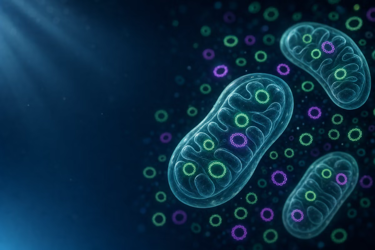
Most of us learn in school that DNA one set of instructions, the same in every cell. But mitochondria quietly break that rule. Each of your cells contains hundreds of mitochondria, and each mitochondrion carries multiple copies of its own small genome, called mitochondrial DNA (mtDNA). That means a single cell might hold thousands of mtDNA molecules altogether.
Here’s where it gets interesting: those thousands of copies don’t have to be identical.
What Is Heteroplasmy?
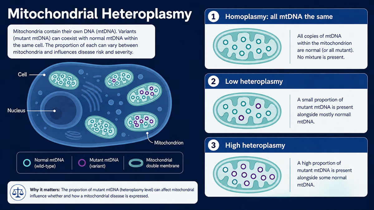
When a mutation arises in one mtDNA molecule, it doesn’t instantly replace all the others. Instead, you can end up with a mixture of some copies carrying the mutation, some perfectly normal. Scientists call this state heteroplasmy (from the Greek for “different” and “formed”). Think of it like a bag of marbles: most are green (normal), but a handful are purple (mutant). The bag is heteroplasmic. If every marble were the same color, that would be homoplasmy, either all normal or all mutant.
Heteroplasmy is the rule rather than the exception in human populations. Studies using sensitive sequencing techniques have found low-level heteroplasmic variants in virtually everyone.
Why Does the Mix Vary?
The proportion of mutant to normal mtDNA isn’t fixed. It can differ dramatically between tissues in the same person. Your muscle cells might carry 70% mutant mtDNA while your blood cells carry only 20%. It can also shift over a lifetime as cells divide and mitochondria replicate, a process called mitotic segregation.
This variability is a big part of why mitochondrial diseases are so hard to predict. Two people with the same mutation can have wildly different proportions of mutant mtDNA with very different symptoms, or none at all. So what does the proportion actually have to do with getting sick? That’s where the threshold effect comes in.
The Threshold Effect
Having some mutant mtDNA in your cells doesn’t automatically mean you’ll develop a disease. Your healthy mitochondria are remarkably good at compensating. As long as enough normal copies are around to keep energy production running, the cell can cope; the mutant copies are essentially outvoted.
Symptoms typically appear only when the proportion of mutant mtDNA climbs above a critical level, often called the pathogenic threshold. For many mtDNA mutations, that threshold sits somewhere between 60% and 90% mutant copies, though the exact number varies by mutation and by tissue.
Think of it like a work team. If ten people are supposed to run a factory and two call in sick, production barely dips. But if eight call in sick, the factory grinds to a halt. The threshold is the point at which there simply aren’t enough healthy workers left to keep things running.
Not all tissues hit that threshold at the same proportion, though. Cells that are especially hungry for energy (neurons in the brain, heart muscle cells, skeletal muscle) have less tolerance for mitochondrial dysfunction and tend to be affected first and most severely. That’s why mitochondrial diseases so often show up as neurological problems, heart conditions, or muscle weakness. The practical takeaway: the same mutation can cause serious disease in one tissue while leaving another completely unaffected.
Maternal Inheritance
Mitochondria are almost exclusively inherited from your mother. When a sperm fertilizes an egg, the egg contributes the cytoplasm, the cellular fluid where mitochondria live, while the sperm contributes almost none. The few paternal mitochondria that do sneak in are typically tagged for destruction by the embryo shortly after fertilization.
This has profound consequences for how mitochondrial diseases are passed down:
- A mother with a pathogenic mtDNA mutation can pass it to all of her children, sons and daughters alike.
- A father with the same mutation cannot pass it on through mtDNA. His children will not inherit it from him.
- An affected son will not pass the disease to his children; an affected daughter may pass it to all of hers.
The Bottleneck Effect
Here’s the twist that explains why siblings can be so differently affected. During the formation of egg cells, the number of mtDNA molecules goes through a dramatic reduction, a genetic bottleneck, before being amplified again. This random sampling means that one egg might end up with a high proportion of mutant mtDNA while another egg from the same mother ends up with very little. The result: children of the same mother can inherit very different heteroplasmy levels, and therefore have very different disease severity or no disease at all.
This bottleneck is one of the most clinically important features of mitochondrial genetics, because it makes predicting disease risk in future children genuinely difficult, even when a mother’s heteroplasmy level is known. Genetic counseling is essential for families navigating this uncertainty.
Why Nuclear-Gene Mitochondrial Disease Exists
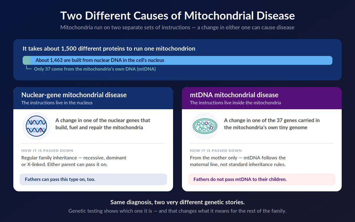
Here’s the surprise twist that catches many people off guard: mitochondria have their own DNA, yes but it’s a very small genome. Human mtDNA encodes only 37 genes. Running a fully functional mitochondrion, however, requires roughly 1,500 proteins. The vast majority of those proteins are encoded not in mtDNA, but in the nuclear genome (the main DNA in the cell’s nucleus) and are imported into the mitochondrion after being made.
This means that mutations in nuclear genes can cripple mitochondrial function just as completely as mutations in mtDNA itself. The affected proteins span a wide range of functions:
- Respiratory chain assembly factors - proteins that build the complexes that generate ATP (your cell’s energy currency)
- mtDNA maintenance proteins - proteins that copy and repair mtDNA itself (mutations here can cause secondary mtDNA deletions or depletion)
- Import machinery proteins - proteins that ferry other proteins across the mitochondrial membranes
- Mitochondrial ribosomal proteins - needed to translate the genes that mtDNA does encode
The critical difference is in how these diseases are inherited. Because nuclear genes follow standard Mendelian rules, nuclear-encoded mitochondrial diseases can be autosomal recessive (two faulty copies needed, one from each parent), autosomal dominant (one faulty copy is enough), or X-linked. None of which show the maternal-only inheritance pattern of mtDNA diseases.
“Mitochondrial disease” is therefore an umbrella term covering two very different genetic architectures: mitochondrial disease caused by a nuclear gene mutation versus mitochondrial disease caused by a mtDNA mutation. Knowing which type you’re dealing with changes everything.
What This Means for You
Mitochondrial genetics can feel overwhelming at first. Mutations that may or may not cause disease, thresholds that differ by tissue, inheritance patterns that depend on whether the mutation is in mtDNA or nuclear DNA. But the science has come a long way, and so have the tools available to families.
Genetic testing today can identify both mtDNA mutations and nuclear-gene causes of mitochondrial disease, often from a blood sample or muscle biopsy. Knowing the specific genetic cause and whether it’s in mtDNA or a nuclear gene is the foundation for understanding inheritance risk, making informed decisions about family planning, and connecting with the right specialists and support communities.
If you or someone in your family has been diagnosed with a mitochondrial condition, or if you’re concerned about symptoms that might point in that direction, a referral to a mitochondrial disease specialist or a clinical geneticist is the best next step. You don’t have to decode this alone.
Frequently Asked Questions
How can you find a clinic or specialist who understands mitochondrial disease and heteroplasmy?
You can start by asking members of your current medical team for a referral to a mitochondrial specialist, since they may already know regional experts or centers. In addition, many patient organizations maintain specialist directories you can search yourself, including the United Mitochondrial Disease Foundation’s “Find a Doctor” tool and MitoAction’s mitochondrial disease doctor listings.
References
[1] Wallace DC. Mitochondrial DNA in aging and disease. Sci Am. 1997;277(2):40-47.
[2] Ye K, Lu J, Ma F, Keinan A, Gu Z. Extensive pathogenicity of mitochondrial heteroplasmy in healthy human individuals. Proc Natl Acad Sci USA. 2014;111(29):10654-10659.
[3] Lightowlers RN, Taylor RW, Turnbull DM. Mutations causing mitochondrial disease: What is new and what challenges remain? Science. 2015;349(6255):1494-1499.
[4] Rossignol R, Faustin B, Rocher C, et al. Mitochondrial threshold effects. Biochem J. 2003;370(Pt 3):751-762.
[5] Chinnery PF, Turnbull DM. Mitochondrial DNA and disease. Lancet. 1999;354 Suppl 1:SI17-21.
[6] Gorman GS, Chinnery PF, DiMauro S, et al. Mitochondrial diseases. Nat Rev Dis Primers. 2016;2:16080.
[7] Sutovsky P, Moreno RD, Ramalho-Santos J, et al. Ubiquitin tag for sperm mitochondria. Nature. 1999;402(6760):371-372.
[8] Cree LM, Samuels DC, de Sousa Lopes SC, et al. A reduction of mitochondrial DNA molecules during embryogenesis explains the rapid segregation of genotypes. Nat Genet. 2008;40(2):249-254.
[9] Steffann J, Gigarel N, Corcos J, et al. Stability of the m.8993T>G mtDNA mutation load during human embryofetal development has implications for the feasibility of prenatal diagnosis in NARP syndrome. J Med Genet. 2007;44(10):664-669.
[10] Calvo SE, Clauser KR, Mootha VK. MitoCarta2.0: an updated inventory of mammalian mitochondrial proteins. Nucleic Acids Res. 2016;44(D1):D1251-D1257.
[11] Vafai SB, Mootha VK. Mitochondrial disorders as windows into an ancient organelle. Nature. 2012;491(7424):374-383.
[12] Chinnery PF. Mitochondrial disease in adults: what’s old and what’s new? EMBO Mol Med. 2015;7(12):1503-1512.